What Do Margins Mean In Breast Cancer / Determination Of Tumor Margins With Surgical Specimen Mapping Using Near Infrared Fluorescence Cancer Research / Tumor margins when breast cancer is surgically removed (during a surgical biopsy, lumpectomy or mastectomy), a rim of normal tissue surrounding the tumor is also removed.
Dapatkan link
Facebook
X
Pinterest
Email
Aplikasi Lainnya
What Do Margins Mean In Breast Cancer / Determination Of Tumor Margins With Surgical Specimen Mapping Using Near Infrared Fluorescence Cancer Research / Tumor margins when breast cancer is surgically removed (during a surgical biopsy, lumpectomy or mastectomy), a rim of normal tissue surrounding the tumor is also removed.. This would be considered a concordant biopsy diagnosis. New evidence about why clear margins in breast cancer surgery are such good news date: The pathologist looks at the margins under a microscope and determines whether or not they contain cancer cells. After the procedure, a pathologist examines the removed tissue to check for remaining cancer cells in the lumpectomy margins. They indicated this is new from my mammo last year.
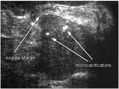
Share your story in our bidmc cancer community. Hearing that your surgeon got clean margins is a cause for relief and gratitude, and means that you can move forward to the rest of breast cancer treatment with optimism. The margin width is the distance from the cancer to the ink painted on the surface of the excision specimen. A breast ultrasound can help in diagnosis in differentiating between benign and malignant tumors, often without the need for a needle biopsy. The risk of breast cancer increases with age.
Infiltrating Ductal Carcinoma Features Of Solid Nodules from breast-cancer.ca Then under diagnosis the report states: The margins of the breast mass are important a radiologist interpreting a breast cancer screening mammogram will be alarmed when they discover a mass with a poorly defined or spiculated margin. If cancer cells are touching the ink (called positive margins), it can mean that some cancer was left behind, and more surgery or other treatments may be needed. Lumpectomy margins are the rim of normal tissue surrounding the cancer tumor that is often removed with the tumor during the surgery to ensure the cancer is completely gone. There is not always agreement on how large of a margin is necessary and sometimes it depends on the type of cancer. They indicated this is new from my mammo last year. In some cases, a pathologist may classify the margins as close, which means that cancer cells are close to the edge of the healthy tissue, but not right at the edge and don't have ink on them. Read more about breast cancer care at bidmc.
The cancer has not spread to distant sites.
When breast cancer is surgically removed (during a surgical biopsy, lumpectomy or mastectomy), a rim of normal tissue surrounding the tumor is also removed. The tumor extends less than 1mm. Margins help show whether or not all of the tumor was removed. If cancer cells are present, this will influence decisions about treatments such as additional surgery and radiation. One of the most important factors associated with local recurrence after lumpectomy in breast cancer patients is the status of the surgical margin. In some cases, a pathologist may classify the margins as close, which means that cancer cells are close to the edge of the healthy tissue, but not right at the edge and don't have ink on them. If your cancer is tricky to diagnose, the pathologist may write extra comments. Then under diagnosis the report states: Standard surgical practice is to obtain clear margins even if this requires a second surgical procedure. The age of the patient and the appearance of the mass with indistinct margins make breast cancer a strong diagnostic possibility. They indicated this is new from my mammo last year. 4 breast infections can cause redness and swelling. Cancer starts in the cells lining the ducts or lobules, when a normal cell becomes a carcinoma cell.
A breast ultrasound can help in diagnosis in differentiating between benign and malignant tumors, often without the need for a needle biopsy. Just got a call from the imaging center that my mammogram showed an oval mass in the lower left breast, middle depth, partially obscured. In some cases, a pathologist may classify the margins as close, which means that cancer cells are close to the edge of the healthy tissue, but not right at the edge and don't have ink on them. New evidence about why clear margins in breast cancer surgery are such good news date: Then under diagnosis the report states:
Resection Margin Wikipedia from upload.wikimedia.org It will usually contain the type of cancer, tumor grade, lymph node status, margin status, and stage. In some cases, a pathologist may classify the margins as close, which means that cancer cells are close to the edge of the healthy tissue, but not right at the edge and don't have ink on them. The age of the patient and the appearance of the mass with indistinct margins make breast cancer a strong diagnostic possibility. The classic description of a breast cancer is a mass with an irregular shape and spiculated margin (fig. Lumpectomy margins are the rim of normal tissue surrounding the cancer tumor that is often removed with the tumor during the surgery to ensure the cancer is completely gone. But not to the deep margin of resection. They're often easy to move around (mobile) and may be tender. It is assumed that reexcision to achieve clear …
Standard surgical practice is to obtain clear margins even if this requires a second surgical procedure.
Margins help show whether or not all of the tumor was removed. Cancer starts in the cells lining the ducts or lobules, when a normal cell becomes a carcinoma cell. This might indicate that breast cancer cells are infiltrating into the surrounding tissue. During or after surgery, a pathologist examines this rim of tissue — called the surgical margin or margin of resection — to be sure it's clear of any cancer cells. I am a 46 y/o female, this was my annual mammogram. A positive margin means that cancer cells come right out to the edge of the removed tissue and have ink on them. The cancer has not spread to distant sites. This would be considered a concordant biopsy diagnosis. There is not always agreement on how large of a margin is necessary and sometimes it depends on the type of cancer. Research shows about 1 out of 4 women who have a lumpectomy go on to have a second breast surgery because the margins weren't clear after their first surgery. The age of the patient and the appearance of the mass with indistinct margins make breast cancer a strong diagnostic possibility. But not to the deep margin of resection. A breast ultrasound can help in diagnosis in differentiating between benign and malignant tumors, often without the need for a needle biopsy.
Standard surgical practice is to obtain clear margins even if this requires a second surgical procedure. The cancer has not spread to distant sites. If cancer cells are touching the ink (called positive margins), it can mean that some cancer was left behind, and more surgery or other treatments may be needed. The classic description of a breast cancer is a mass with an irregular shape and spiculated margin (fig. During or after surgery, a pathologist examines this rim of tissue — called the surgical margin or margin of resection — to be sure it's clear of any cancer cells.
Not All Breast Cancers Are The Same from post.healthline.com The tumor extends less than 1mm. Margins more widely clear than 2 mm do not further reduce the rates of recurrence of cancer in the breast and their routine use is not supported by evidence. A breast ultrasound can help in diagnosis in differentiating between benign and malignant tumors, often without the need for a needle biopsy. Read more about breast cancer care at bidmc. There is a tumor in the breast that is ≤2 cm. After the procedure, a pathologist examines the removed tissue to check for remaining cancer cells in the lumpectomy margins. The classic description of a breast cancer is a mass with an irregular shape and spiculated margin (fig. The pathologist looks at the margins under a microscope and determines whether or not they contain cancer cells.
After the procedure, a pathologist examines the removed tissue to check for remaining cancer cells in the lumpectomy margins.
One of the most important factors associated with local recurrence after lumpectomy in breast cancer patients is the status of the surgical margin. In some cases, a pathologist may classify the margins as close, which means that cancer cells are close to the edge of the healthy tissue, but not right at the edge and don't have ink on them. Clear margins are associated with a lower risk of a local recurrence (cancer returning in the same breast). Research shows about 1 out of 4 women who have a lumpectomy go on to have a second breast surgery because the margins weren't clear after their first surgery. This rim is called a margin. The tumor extends less than 1mm. Clinical judgment should be used in determining the need for further surgery in patients with negative margins less than 2 mm. It is assumed that reexcision to achieve clear … The margin width is the distance from the cancer to the ink painted on the surface of the excision specimen. Hearing that your surgeon got clean margins is a cause for relief and gratitude, and means that you can move forward to the rest of breast cancer treatment with optimism. This would be considered a concordant biopsy diagnosis. When breast cancer is surgically removed (during a surgical biopsy, lumpectomy or mastectomy), a rim of normal tissue surrounding the tumor is also removed. This rim is called a margin.
17.11.2021 · reuters logos and exchange rates of bitcoin, litecoin, monero and ether to swiss franc are seen on a cryptocurrency atm in zurich. Simply enter your business name and make a logo you'll love. Bitcoin falls more than 4% to near $60,000 ; The cryptocurrency cart continued to bleed on … Metaverse and its connection with cryptocurrency; What is a Cryptocurrency? â€" CriptoMonedas from criptomonedas.org The cryptocurrency cart continued to bleed on … Simply enter your business name and make a logo you'll love. 17.11.2021 · reuters logos and exchange rates of bitcoin, litecoin, monero and ether to swiss franc are seen on a cryptocurrency atm in zurich. Bitcoin falls more than 4% to near $60,000 ; Crypto turns rs 1,000 into rs 2.37 crore in a day; Metaverse and its connection with cryptocurrency; Browse stunning logos ta...
How To Make My Phone Read My Sd Card : Can T Write To Sd Card On Samsung Phone How To Fix - Press the blackberry menu button and select cut.. . Check if sd card reader is defective. A temporary workaround would be to connect the camera, phone, or another device to the computer using a usb cable with the sd card in the device. Encryption converts data from a readable form to an encoded version that can only be read on the device that was used to encrypt it. Here, expand the memory technology devices section and check if there's an issue sign next to your card reader. Select device and navigate to camera.. Check if you phone or computer is able to read the sd card. Plug one end of the usb cable that comes with your phone to the mini usb port in your phone and plug the other end to a usb port in your computer. First unplug the sd card reader from your phone or computer. Want to transfer music to sd but can't find where it's stored internally: Check if sd c...
How To Make A Card For Mother's Day : 8 Mother S Day Cards The Kids Can Make Studio 5 : With patterned paper, a few embellishments, and a little creativity, you can create a use this free mother's day card printable to tell mom how you feel. . On paper or wallpaper with a pattern cut rectangle is sized postcards page, and then stick it inside (if you have a color printer, you can print a picture template located below). 30 crafty homemade diy mother's day card ideas if you're wondering how to make a mother's day card. Glue the strips in the top right cards right, then. This makes one of the best homemade mother's day cards for kids to make. Show your affection with a heartfelt, handmade mother's day card featuring everlasting flowers. With social media and text messaging, it's easy to throw her a text or tag her in your status to send your greetings. If you want to make mother's day 2021 extra memorable, you can't go w...


Komentar
Posting Komentar